Helicosporidia project online
Home
Helicosporidia
Photo Gallery
Cyst Gallery
Vegetative cell Gallery
Harpoon cell Gallery
Pellicle Gallery
In vivo development gallery
In vitro development gallery
Methodology
Molecular Biology
People
Publications
Links

In vivo gallery | 1 | 2 | 3 | 4 | Select Gallery

Vegetative growth (con't)

Light micrograph of hemolymph sampled from Helicosporidium spp.-infected Spodoptera exigua larvae 96 hours after cyst ingestion. Multiple vegetative cells being released from a cluster of hemocytes.

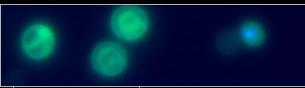
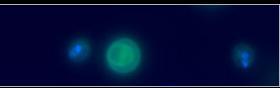
Light micrograph of hemolymph sampled from Helicosporidium spp.-infected Spodoptera exigua larvae 168 hours after cyst ingestion. (H) Several freely circulating vegtative cells, pellicles, and a few cysts. (I) Massive numbers of freely-circulating cysts and vegetative cells. Cyst development also occurs within hemocytes. (Bar = 10 µm)

In vivo gallery | 1 | 2 | 3 | 4 | Select Gallery