Helicosporidia project online
Home
Helicosporidia
Photo Gallery
Cyst Gallery
Vegetative cell Gallery
Harpoon cell Gallery
Pellicle Gallery
In vivo development gallery
In vitro development gallery
Methodology
Molecular Biology
People
Publications
Links

Harpoon Cell Gallery | Select Gallery

Light Microscopy

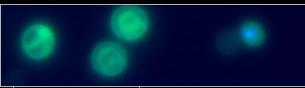
Preparation where cysts have opened or dehisced releasing the filamentous cell-ovoid cell complex from the pellicle. The pressure applied to the coverslip results in a fracturing of the pellicle, a swelling of the central ovoid cells, and an uncoiling of the filamentous cells. In several cases, the three ovoid cells and the filamentous cells can be observed in proximity with the pellicle.

SEM

SEM of the midgut region of Manduca sexta at 4 h post-challenge with Helicosporidium sp. Filamentous cells have penetrated the microvillar lining of these midgut cells.

A filamentous cell embedded in a midgut cell demonstrating the orientation of the surface projections.

SEM of dehisced helicosporidial cyst. The uncoiled filamentous cell extending away from the ovoid cell complex.

Harpoon Cell Gallery | Select Gallery