|
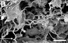
Scanning electron micrograph of the midgut lumen of Spodoptera exigua larvae after oral challenge with Helicosporidiumspp. cysts. Note numerous cysts and filaments in the food bolus 8 hours after ingestion. (Bar = 10 µm) |
|
Scanning electron micrograph of the midgut lumen of Spodoptera exigua larvae after oral challenge with Helicosporidiumspp. cysts. Four hours after ingestion. (Bar = 10 µm) |
|
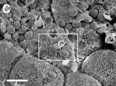
Intact and dehiscing cysts in association with the microvillar lining of the midgut epithelium 4 hours after ingestion. (Bar = 10 µm) |
|
Intact and dehiscing cysts in association with the microvillar lining of the midgut epithelium 4 hours after ingestion. (Bar = 3 µm) |
|
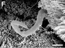
A filament entering the midgut epithelium 24 hours after cyst ingestion. (Bar = 10 µm) |
|
Note orientation of characteristic barbs, pointing towards the midgut lumen. (Bar = 1 µm) |