|
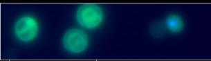
Light micrograph of hemolymph sampled from Helicosporidium spp.-infected Spodoptera exigua larvae 48 hours after cyst ingestion. Two hemocytes, one containing a bean-shaped vegetative cell. |
|
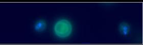
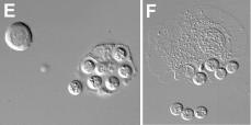
Light micrograph of hemolymph sampled from Helicosporidium spp.-infected Spodoptera exigua larvae 48 hours after cyst ingestion. A quartet of early vegetative cells within a hemocyte. |

|
Light micrograph of hemolymph sampled from Helicosporidium spp.-infected Spodoptera exigua larvae 48 hours after cyst ingestion. Clustered hemocytes containing vegetative cells (black arrows) and the remnants of a filament (white arrow). Note the cell division within the filamentous cell. |
Vegetative growth |

|
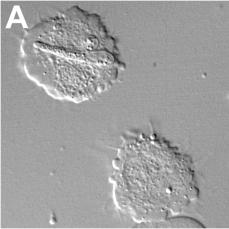
Light micrograph of hemolymph sampled from Helicosporidium spp.-infected Spodoptera exigua larvae 72 hours after cyst ingestion. Two hemocytes, one containing an octet of vegetative cells and displaying cytopathic effects, the other containing a bean-shaped vegetative cell. |
|
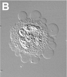
Light micrograph of hemolymph sampled from Helicosporidium spp.-infected Spodoptera exigua larvae 72 hours after cyst ingestion. Hemocytes releasing vegetative cells. |