|
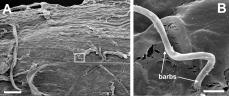
Scanning electron micrograph of basal midgut surface of Spodoptera exigua larvae showing invasive filaments gaining ingress into the hemocoel 8 hours after ingestion of spp. cysts. (Bar = 30 µm). At right, note the orientation of barbs toward the hemocoel (Bar = 3 µm). |
|
Scanning electron micrograph of basal midgut surface of Spodoptera exigua larvae showing invasive filaments gaining ingress into the hemocoel 8 hours after ingestion of spp. cysts. (Bar = 30 µm). At right, note the orientation of barbs toward the hemocoel (Bar = 1 µm). |

|
Scanning electron micrograph of basal midgut surface of Spodoptera exigua larvae showing invasive filaments gaining ingress into the hemocoel 12 hours after ingestion of spp. cysts. (Bar = 100 µm). At right, note the orientation of barbs toward the hemocoel (Bar = 3 µm). |

|
Scanning electron micrograph of basal midgut surface of Spodoptera exigua larvae showing invasive filaments gaining ingress into the hemocoel 24 hours after ingestion of spp. cysts. (Bar = 30 µm). At right, note the orientation of barbs toward the hemocoel (Bar = 3 µm). |

|
Scanning electron micrographs of the basal midgut surface of Spodoptera exigua larvae 12 hours after cyst ingestion, showing a filament partially engulfed by a phagocytic hemocyte. (Bar at left = 30 µm; Bar at right = 3 µm) |