|
|
Cyst preparation and DNA extraction
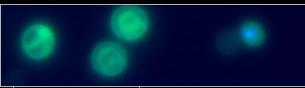
Helicosporidium sp.
was isolated originally from the blackfy Simulium jonesi
Stone & Snoddy (Diptera: Simuliidae) and produced in
Helicoverpa zea. Approximately 4e7 cysts suspended in
0.15 M NaCl were applied to a linear gradient of 1.00-1.3003 g Ludox HS40 ml-1 (DuPont). Helicosporidial cysts
that banded at an estimated density of 1.17 g ml-1 were
collected, diluted in 10 vols deionized water and washed free
of residual Ludox by repeated low-speed centrifugation
steps. The pellet, resuspended in 50 µl water, was extracted
with the use of the Masterpure Yeast DNA extraction kit
(Epicentre Technologies), following the manufacturer's protocol.
Examination of the cells before and after lysis
treatment revealed the presence of numerous, highly
refractile cysts before treatment and, after incubation in the
lysis buffer at 50°C, cysts appeared to dehisce, releasing the
filamentous cells. However, no massive disruption of the
ovoid cells or filamentous cells was observed in these
preparations. Visible pellets were observed after RNase
treatment, phenol/chloroform extraction and ethanol precipitation.
The final pellet, suspended in molecular-biology-grade
water, was frozen at -20°C. The DNA preparations,
electrophoresed on a 0.8% agarose gel and stained with
ethidium bromide, produced a single, discrete ~20 kb band
diagnostic for genomic DNA.
Mature cysts were also processed for electron microscopy
according to previously published protocols (Boucias et al.,
2001).
|