|
|
Helicosporidium Infection Experiments | 1 | 2 | 3
Printable version
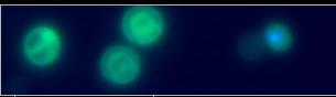
Helicosporidium:Cysts were amplified in vivo in orally infected H. zea larvae and purified from homogenates of late instars and pupae by high-speed centrifugation on a linear gradient of Ludox HS40 (DuPond Chemical, Boston, MA). The band containing the cysts was collected, diluted in sterile water, and subjected to several cycles of low-speed centrifugation to remove residual gradient material. After re-suspension in sterile water or in 10% DMSO plus 10% glycerol for cryo-protection, cysts were counted using a hemacytometer and aliquots with a concentration of 1x10⁸ cysts/ml were stored at -70 °C until used. Prior to each experiment, cysts were thawed quickly, washed twice in sterile water, and re-suspended in 0.1 % aqueous Nile Blue A or in sterile water.
Insects: Larvae of H. zea and eggs of S. exugia and T. ni were obtained from established colonies at the Center for Medical, Agricultural and Veterinary Entomology, USDA, ARS, Gainesville, Florida. Hatching neonates and larvae were provided with artificial insect diet and maintained at constant conditions (27 ± 1 °C, 70% RH, 12-h photoperiod). Third instars were used for oral treatments; injection treatments were conducted using fifth instars.
Helicosporidium Infection Experiments | 1 | 2 | 3
|