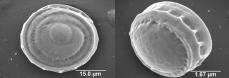
Narrow surface of a cyst depicting the coiled filament cell underlying the pellicle, and the broad surface of a cyst.
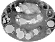
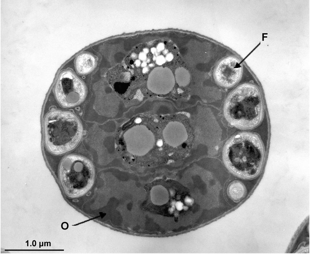
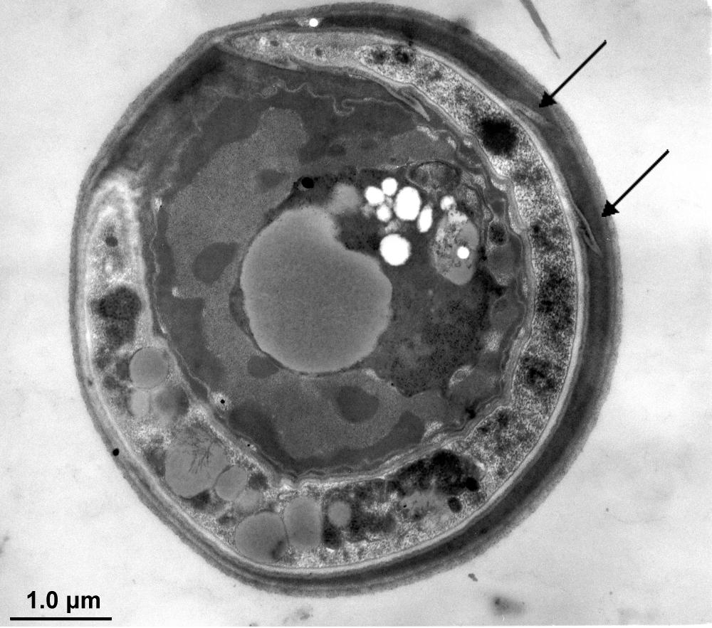
Transmission electron micrographs of the cysts of Helicosporidium sp. from the black fly Simulium jonesi.
Mature cyst composed of three central ovoid cells (O) and the peripherally located filamentous cell (F) contained within a multi-layered cyst wall.
Sagittal section of the filamentous cell within the cyst demonstrating several of the projections (arrows) on the cell wall.