|
|
Studies on In Vivo Development of Helicosporidium
In vivo | 1 | 2
Printable version
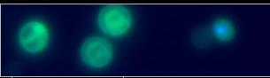
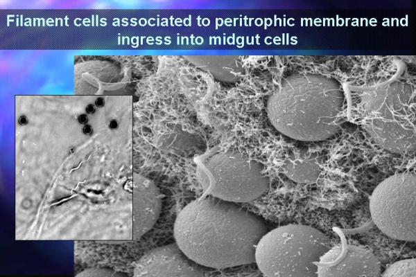
The two Helicosporidia isolates are capable of infecting and replicating in a variety of dipteran and lepidopteran species. Oral challenge of H. zea and the tobacco hornworm M. sexta larvae with cyst preparations was lethal to tested insects. Examination of dissected alimentary tracts revealed that ingested cysts bound initially to the peritrophic matrix in challenged larvae. Within 2 h after ingestion, cysts dehisced releasing filamentous cells from the ovoid cell-pellicle complex. SEM of the midguts dissected from Manduca sexta larvae at 4 h after ingestion revealed that the released filamentous cells penetrated the peritrophic matrix and attached to the midgut columnar epithelium  . Filamentous cells penetrated the midgut with the projections oriented away from the penetration point, suggesting that these spines may play a role in anchoring the filamentous cell to the gut epithelium. In the case of M. sexta, vegetative cells were observed in the hemolymph within 2 days after ingestion. Vegetative cells, containing variable numbers of cells within the pellicles, were observed to be both associated with circulating hemocytes and present as freely circulating cells . Filamentous cells penetrated the midgut with the projections oriented away from the penetration point, suggesting that these spines may play a role in anchoring the filamentous cell to the gut epithelium. In the case of M. sexta, vegetative cells were observed in the hemolymph within 2 days after ingestion. Vegetative cells, containing variable numbers of cells within the pellicles, were observed to be both associated with circulating hemocytes and present as freely circulating cells  . By 6 days post ingestion plasmodial-like hemocytes displayed marked cytopathic effects (CPE). It is unclear whether the Helicosporidium sp. induced a haemocyte fusion, blocked cytokinesis, or stimulated hemocyte nuclear division. Within 10-14 days, treated larvae contained massive numbers of mature cysts in the cream-colored hemolymph . By 6 days post ingestion plasmodial-like hemocytes displayed marked cytopathic effects (CPE). It is unclear whether the Helicosporidium sp. induced a haemocyte fusion, blocked cytokinesis, or stimulated hemocyte nuclear division. Within 10-14 days, treated larvae contained massive numbers of mature cysts in the cream-colored hemolymph  . At this point large numbers of cysts could be extracted easily using several cycles of centrifugation followed by high-speed centrifugation through a Ludox gradient. . At this point large numbers of cysts could be extracted easily using several cycles of centrifugation followed by high-speed centrifugation through a Ludox gradient.
In vivo | 1 | 2
|