|
|
Studies on In Vivo Development of Helicosporidium
In vivo | 1 | 2
Printable version
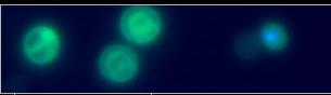
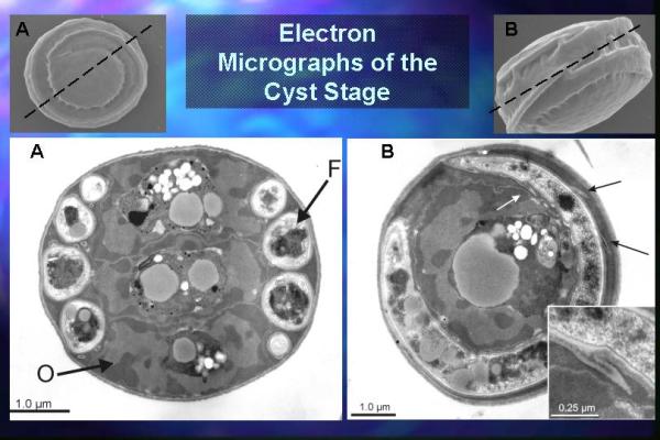
Recently, Helicosporidium has been isolated from larvae of the black fly Simulium jonesi Stone & Snoddy (Diptera, Simuliidae) collected in 1998 from Hatchet Creek, Alachua Co, Fl. and from the weevil Cyrtobagous salviniae Calder & Sands (Coleoptera: Curculionidae) also found in Gainesville, Florida. This insect is an introduced biological control agent for the aquatic weed Salvinia molesta (Salviniaceae). Phase contrast microscopy of tissue smears revealed discoid cysts that measured 6.5 ± 0.2 x 5.9 ± 0.3 µm. Light and electron microscope studies demonstrated that the cysts contained a core of three ovoid cells and a single filamentous cell.  . The filamentous cell makes 3-4 coils, forming a helix around the ovoid cells within the mature cysts. The outer wall or pellicle is a multilaminate structure, enclosing the peripheral filamentous cell within its innermost wall layer. Within intact cysts, three centrally located ovoid cells are compressed in an accordion-like fashion. Each of these cells possesses a peripheral nucleus that encloses a cytoplasmic region that contain a variety of vacuoles and granules. . The filamentous cell makes 3-4 coils, forming a helix around the ovoid cells within the mature cysts. The outer wall or pellicle is a multilaminate structure, enclosing the peripheral filamentous cell within its innermost wall layer. Within intact cysts, three centrally located ovoid cells are compressed in an accordion-like fashion. Each of these cells possesses a peripheral nucleus that encloses a cytoplasmic region that contain a variety of vacuoles and granules.
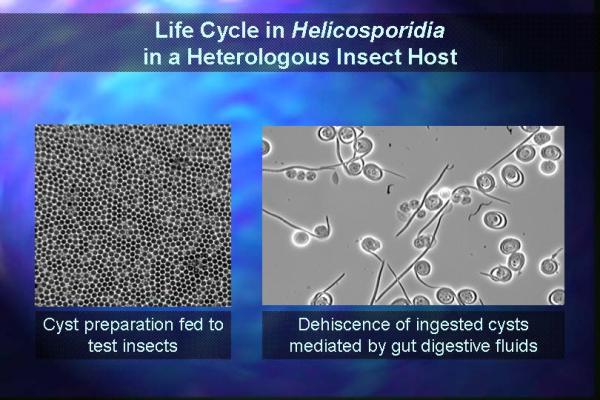
When stimulated by pressure, the outer pellicle layer of the cyst splits open or dehisces releasing the filamentous cell-ovoid cell complex from the pellicle  . Release from the cyst stage produces an expanded ovoid cell aggregate and results in the uncoiling of filamentous cells. The filamentous cells, measuring 37 ± 4.3 mm in length by 0.9 ± 0.13 mm in diameter, are coated with short projections (340 ± 60 nm) orientated in the same direction providing polarity to the filamentous cells . Release from the cyst stage produces an expanded ovoid cell aggregate and results in the uncoiling of filamentous cells. The filamentous cells, measuring 37 ± 4.3 mm in length by 0.9 ± 0.13 mm in diameter, are coated with short projections (340 ± 60 nm) orientated in the same direction providing polarity to the filamentous cells  . Cyst dehiscence was triggered readily by the application of gentle pressure to the coverslip covering a cyst suspension. Alternatively, the incubation of purified cysts in midgut fluid extracted from Heliocoverpa zea larvae stimulated the release of the filamentous cells from both cyst suspensions. A twenty minute exposure to midgut fluids resulted in more than 50% of the cysts releasing their filamentous cells. Upon activation these cysts increased in volume resulting in pellicle rupture and release of the filamentous cell. Incubated in the midgut luminal fluid the ovoid cells lysed, whereas the released filamentous cell became uncoiled and remained intact. Presently, the component(s) in the midgut fluid that signals dehiscence is not known. Uncoiled filamentous cells of the blackfly isolate readily clustered with other filamentous cells producing rosettes. Whether this clumping was due to a specific surface adhesion or was simply a result of entanglement of the surface projections is unknown. The filamentous cells of the weevil isolate did not aggregate when released from the cysts. . Cyst dehiscence was triggered readily by the application of gentle pressure to the coverslip covering a cyst suspension. Alternatively, the incubation of purified cysts in midgut fluid extracted from Heliocoverpa zea larvae stimulated the release of the filamentous cells from both cyst suspensions. A twenty minute exposure to midgut fluids resulted in more than 50% of the cysts releasing their filamentous cells. Upon activation these cysts increased in volume resulting in pellicle rupture and release of the filamentous cell. Incubated in the midgut luminal fluid the ovoid cells lysed, whereas the released filamentous cell became uncoiled and remained intact. Presently, the component(s) in the midgut fluid that signals dehiscence is not known. Uncoiled filamentous cells of the blackfly isolate readily clustered with other filamentous cells producing rosettes. Whether this clumping was due to a specific surface adhesion or was simply a result of entanglement of the surface projections is unknown. The filamentous cells of the weevil isolate did not aggregate when released from the cysts.
In vivo | 1 | 2
|