|
|
Introduction | 1 | 2 | 3
Printable version
Following the
discovery of another isolate of Helicosporidium
parasiticum in a larva of Hepialis
pallens (Hepialidae, Lepidoptera), another taxonomic position was proposed
for the group Helicosporidia. Based on observation of this new isolate as well
as the original specimen described by Keilin, Weiser (1970) claimed that the
Helicosporidia are best placed among the lower Fungi. He argued that the spore
characteristics are too much different from what is found in Protozoa, but are
similar in some aspects to primitive Fungi, such as insect pathogens of the
genus Monosporella, classified as
Nematosporoideae inside the Saccharomycetaceae (primitive Ascomycetes).
In the ealy 1970s Helicosporidium
parasiticum was isolated from larvae and adults of the beetle Carpophilus
mutilatus Erichson (Nitidulidae, Coleoptera). This isolate was infectious per
os to 18 species of arthropods, including three orders of insects
(Lepidoptera, Coleoptera, Diptera) and one family of mites (Kellen and Lindegren 1973). Contrarily, species
of Orthoptera, Hymenoptera and Diptera were not susceptible to this isolates.
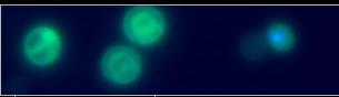
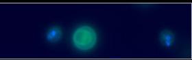
The spores (cysts), present in the host artificial diet, were ingested and
released the three round cells and the filamentous cells in the host midgut.
After 24 h, helicosporidial cells appeared in the host hemolymph and grew
vegetatively. The vegetative growth is characterized by cell division that
occurs within a pellicle. After division, the pellicle ruptures and releases the
daughter cells (4 or 8). Empty pellicles and daughter cells eventually fill the
entire host hemocoel. Daughter cells then develop into spores, in which the
filamentous cell differentiates and encircles the three round cells. Additional
studies reported the presence of Helicosporidium
sp. in new host species, such as crustaceans, mites and collembola, and
trematodes.
Introduction | 1 | 2 | 3
|