|
|
Helicosporidium Infection Experiments | 1 | 2 | 3
Printable version
Mating Experiments:
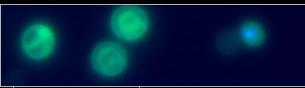
To examine adult fecundity and vertical transmission of the pathogen, mating experiments were conducted with infected and control adults obtained from injection treatments with S. exigua. One female and one male were transferred into a 16-oz-paper cup. A piece of tissue towel along the edge served as egg laying substrate and a Kimwipe® soaked in a 10-% sucrose solution in a small Petri dish (60 mm diameter) served as nutrient source. Each cup was held in a separate Perspex box to prevent pheromone interference between different mating trials. Insects were incubated at constant conditions (27±1 °C, 70 % RH, 12-h photoperiod) and the numbers of deposited eggs were recorded daily. When eggs were laid, pieces of artificial diet were added to the cup and humidity was increased by covering the bottom of the Perspex box with wet paper towels. Dead adults were removed from the cup and their infection status was confirmed by examination of tissue smears for the presence of Helicosporidia using light microscopic optics with differential interference contrast (Leica DM R, Leica Microsystems Wetzlar GmbH, Wetzlar, Germany). Two to three days after egg hatch, 50 second instars were transferred to a clean paper cup with artificial diet and allowed to undergo metamorphosis. Upon pupation, the infection status of 25 pupae was determined as described for the infection bioassays. During the course of the experiment, smears of 10 second instars and hemolymph samples of 20 fourth instars, taken from the original mating arena, were also examined for the presence of Helicosporidia
Helicosporidium Infection Experiments | 1 | 2 | 3
|