University of Florida

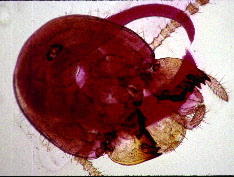
Head of a termite parasitized by the nematode
Description males , Females , Infective juveniles , Type host , Bionomics , Biocontrol , Morphometrics

Head of a termite parasitized by the nematode
Description males , Females , Infective juveniles , Type host , Bionomics , Biocontrol , Morphometrics
No second generation males were found.

Females: (Fig.
2) Anterior region similar to male but much larger. Body spiral,
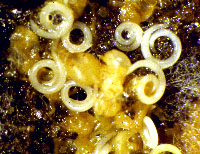
usually found in surrounding medium next to termite cadavers. Cuticle annulated.
Lateral fields not observed. Head truncate or rounded. Labial papillae
six, prominent, many additional papillae-like structures often present
together with labial papillae. Stoma as in male but structures larger and
more prominent; raised ring prominent in stoma apearing tooth-like in lateral
view. Esophagus typical of steinernematids. Gonads didelphic, amphidelphic,
reflexed. Epiptygma absent.Tail curved, longer than body width at anus,
tail length divided by body width at anus about 1.40 (1.10-1.70) (FEMALE.SEM)
. Phasmids prominent, on a protuberance, located in posterior half
of tail. Eggs are not laid but hatch inside the body, and the juveniles
become infective stage before exiting the female.

Females emerged from termites
No second generation females were found.
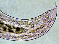
Infective juveniles: (Fig. 3) Juveniles become infective stage juveniles before emerging from female. Body thin, elongate (a=39), well striated. Sheath (second stage cuticle) present but sometimes lost.Head region smooth, mostly slightly enlarged. Oral aperture closed, triradiate; lips six. Labial papillae not observed; four cephalic papillae prominent, situated well behind anterior end. Amphids large, slit like, post-labial in position but anterior to cephalic papillae. Esophagus degenerate, basal bulb elongate with valve, esophago-intestinal valve present but sometimes obscure. Excretory pore prominent; excretory duct long, sinuous, seen clearly on living specimens. A sclerotized structure associated with excretory duct just anterior to excretory cell, appearing as two black dots in lateral view. Lateral field at mid body wider than one third of body width, usually only two border striae seen clearly; eight smooth bands and nine incisures prominent in anterior and posterior regions. Tail elongate, 10 times (8-11) (Table 1) as long as body width at anus, always curved ventrally in posterior end. Average tail length as long as esophagus length. Phasmids prominent, slit-like (JUVENILE.SEM). Left and right phasmids not at the same level, usually situated 25 to 50 m posterior to anus, and occupying 3 or 4 lateral bands.
DISTRIBUTION AND HOSTS
Neosteinernema longicurvicauda has been reported only from the termite Reticulitermes flavipes in Florida, the United States.
BIONOMICS AND HOST PARASITE RELATIONSHIPS
The bionomics and host relationships were report briefly by Nguyen and Smart, 1994. The nematode developed only in the head of the termite. The adults emerged when the tissue of the termite began to decay. The number of adults in each termite was reported as from 1-4 (26% with one nematode, 40 % with two, 20 % with three and 14 % with 4). Twenty of 35 termites studied harbored both males and females, and 15 harbored either males or females. Where only one sex was present, no reproduction occured. When both males and females were present, the females, after emerging, moved a short distance away from the termite cadavers, assumeed a spiral shape, and became immobile. The female retained all eggs that hatched, and the juveniles underwent two molts to become infective juveniles. When most of the juveniles became infective juveniles, they moved vigorously, broke through and emerged from the then female cadaver in the region of tail or stoma. No first- and second-stage juveniles were observed in or surrounding termite cadavers. During developement of juvenile, large number of bacterial cells were observed in the female bodies. No second generation was observed.
| Character1 | infective juvenile(50) | female(25) | male(25) |
|---|---|---|---|
| Body length | 92 SD 60 (789-1,084) | 3,444 SD 784 (2,250-4,766) | 1,236 SD 228 (854-1,084) |
| Greatest width | 24 SD 2 (20-31) | 177 SD 37 (122-250) | 97 SD 18 (67-140) |
| Stoma length | - | 8 SD 1.3 (6-11) | 5.5 SD 1 (5-8) |
| Stoma width | - | 14 SD 1.6 (11-17) | 7.2 SD 1.4 (5-11) |
| EP | 68 SD 4 (61-76) | 112 SD 16 (84-147) | 82 SD 2 (59-103) |
| EPW | 18 SD 2 (10-20) | - | 44 SD 5 (33-52) |
| NR | 107 SD 7 (92-125) | 177 SD 21 (150-225) | 126 SD 15 (103-156) |
| ES | 164 SD 10 (144-188) | 262 SD 28 (219-334) | 187 SD 22 (147-227) |
| Testis flexure | - | - | 277 SD 55 (178-394) |
| Tail length (T) | 167 SD 11 (141-190) | 105 SD 12 (88-134) | 48 SD 6 (36-63) |
| ABW | 17 SD 1 (14-20) | 76 SD 10 (63-103) | 60 SD 10 (41-78) |
| T/ABW | 10 SD 1 (8-11) | 1.4 SD 0.1 (1.1-1.7) | |
| Digitate part of tail | - | 8 SD 3 (3-14) | |
| Spicule length (SP) | - | - | 61 SD 4 (52-67) |
| Spicule width | - | - | 13 SD 1 (11-15) |
| Gubern length (GU) | - | - | 9 SD 4 (52-66) |
| Gubern width | - | - | 9 SD 1 (8-13) |
| Vulva % | - | 55 SD 3 (49-60) | |
| a | 39 SD 3 (30-46) | ||
| b | 5.6 SD 5 (5-7) | ||
| c | 5.5 SD 0.3 (4.7-6.5) | ||
| D%=EP:ES x 100 | 41 SD 2 (38-46) | 43 SD 7 (31-64) 44 SD 6 (30-54) | |
| E%=EP/T x 100 | 41 SD 3 (37-48) | ||
| SW=SP/ABW | - | - | 1.03 SD 0.18 (0.8-1.5) |
| GS=GU/SP | - | - | 0.97 SD 0.06 (0.84-1.08) |