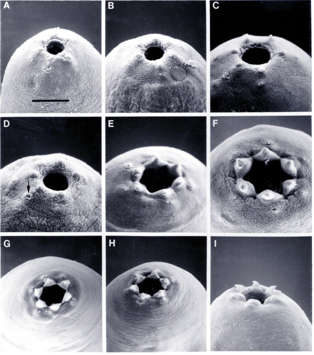
Fig. 3. SEM photographs of Heterorhabditis, day three after infection. A-C) Different face views H. bacteriophora J4s showing round oral aperture, low lips, and labial papillae. The circular structure in B is unknown, seen on this specimen only. D-F) H. megidis. D) Face view of J4 showing round oral aperture, six labial papillae and one of the two amphids (arrow). E, F) Face views of young hermaphrodites showing amphids, elevated lips and labial papillae, and hexagonal-shaped oral aperture. G-I) Different face views of H. hawaiiensis young hermaphrodites showing elevated and outward curved lips and labial papillae, amphids and hexagonal oral aperture. All magnifications based on scale bar in A: A = 5 um, B = 6 um, C = 3.5 um, D = 4.3 um, E = 7.5 um, F = 6.1 um, G,H = 10 um, I = 8.6 um.