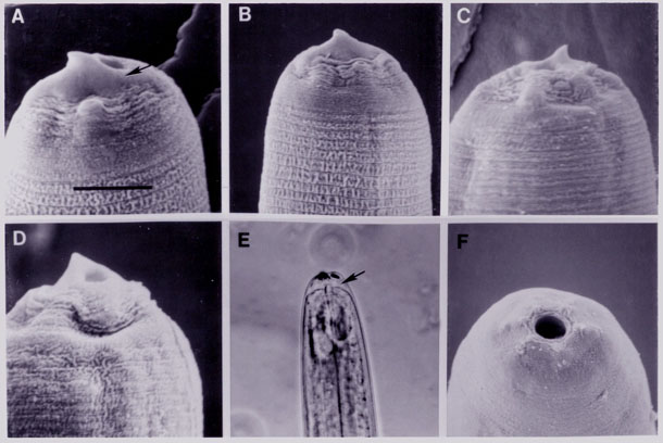
Fig. 2. SEM photographs of Heterorhabditis, day one and day two after
infection. A) Lateral view of the
anterior region of H. megidis J3 at day one showing oral aperture,
dorsal tooth, thickened base of the tooth
(arrow) and amphid. (B) Same as A except dorsal view. C) Anterior region
of H. hawaiiensis J3 at day one
showing oral aperture and dorsal tooth. D) Anterior region of H. megidis
J3 at day two showing dorsal tooth
and collapsed head. E) Light microscope photograph of H. megidis anterior
end, taken at the same time as the
SEM photograph in D, showing space between cuticles in anterior end
(arrow) when molting process begins.
F) Anterior end H. hawaiiensis J4 at day two showing truncated head
with round oral aperture and
inconspicuous lips and labial papillae. All magnifications based on
scale bar in A: A = 3.8 um, B = 5 um, C =
4.3 um, D = 3.8 um, E = 14.5 um, F = 7.5 um.