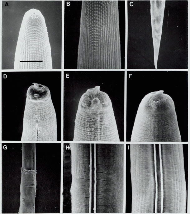
Fig. 1. SEM photographs of Heterorhabditis spp. IJs. A-C) IJ of H. megidis in the J2 cuticle (sheath) showing labial region, tessellate pattern anteriorly, and longitudinal ridges which extend almost the entire length of the body. D, E) anterior region OF H. megidis IJ without the j2 cuticle showing dorsal tooth, membranous ring around the mouth, amphid aperture, beginning line of lateral field and annulation. F) H. bacteriophora anterior region. G) H. hawaiiensis body with anterior part of J2 cuticle (sheath) shed showing two ridges in lateral field. H, I) H. bacteriophora and H. megidis, respectively, showing two ridges in lateral field and annulation. All magnifications based on scale bar in A: A = 8.6 um, B = 10 um, C = 23.1 um, D = 6 um, E = 4.3 um, F = 3.8 um, G = 20 um, H = 5 um, I = 7.5 um.