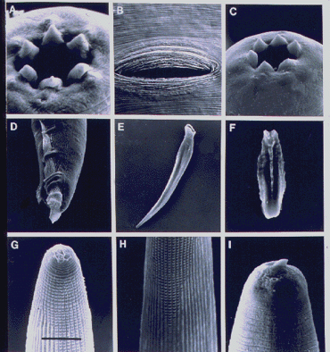
Diagnosis: Hermaphroditic female (Fig. A,B): After entry into an insect host, infective juveniles developing into hermaphroditic females. Head truncate to slightly rounded, six conical lips well developed (Fig. A), separate, each with a terminal papilla; one or two small raised structures sometimes visible at the base of each lip; amphidial opening small. Stoma wide but shallow; cheilorhabdions present, forming a ring, in lateral view resembling two refractile dots. Other parts of the stoma fused to form a collapsed posterior portion. Posterior part of stoma covered by esophagus. Esophagus without metacorpus; isthmus slender; basal bulb swollen; valve in basal bulb reduced. Nerve ring at middle of isthmus. Excretory pore usually posterior to end of esophagus. Vulva median, slit-like, surrounded by elliptical rings (Fig. B); ovotestis amphidelphic, reflexed. Oviparous, later becoming ovoviviparous. Tail pointed, longer than anal body width, postanal swelling usually present.
Amphimictic females (Fig. C): Similar to, but usually smaller than, hermaphroditic females; labial papillae prominent. Reproductive system amphidelphic, vulva not functional for egg deposition, but functional for mating.
Males (Fig. D-F): Testis one, reflexed. Spicules paired, separate, slightly curved ventrally (Fig. E). Spicule head short, offset from lamina by a constriction. Gubernaculum (Fig. F) usually about half as long as spicule length. Bursa peloderan (Fig. D) with nine pairs of genital papillae.
Infective juveniles (Fig.
G-I): Third-stage infective juvenile (IJ) usually with sheath (cuticle
of second-stage juvenile). Sheath with anterior tessellate pattern (Fig.
G) and longitudinal ridges (Fig. H); IJ cuticle striated with one smooth
band marginated by two ridges in lateral fields. Head with prominent dorsal
tooth (Fig. I). Mouth and anus closed. Stoma appearing as a closed chamber
with parallel walls. Esophagus and intestine reduced. Excretory pore posterior
to nerve ring. Symbiotic bacterial cells found in intestine. Tail pointed.
Type species: Heterorhabditis bacteriophora Poinar, 1976