common name: velvet ants
scientific name: Mutillidae (Insecta: Hymenoptera)
Introduction - Distribution - Biology - Identification - Key to Florida Genera - Florida Species - Pest Status and Management - Acknowledgments - Selected References
Introduction (Back to Top)
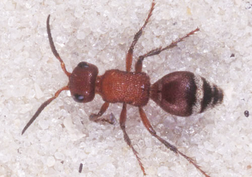
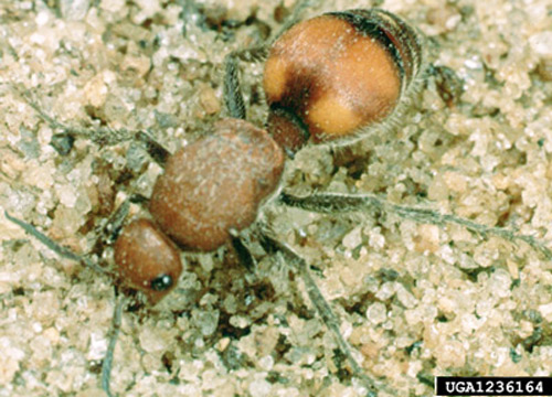
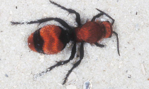
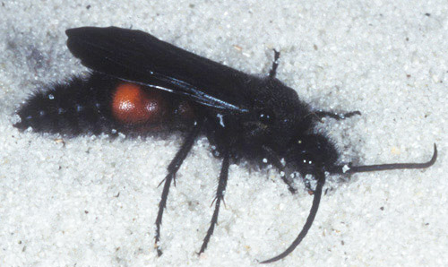
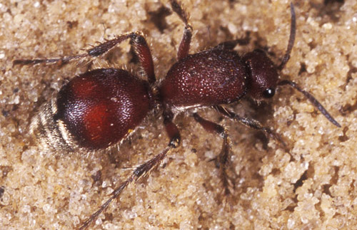
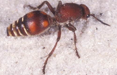
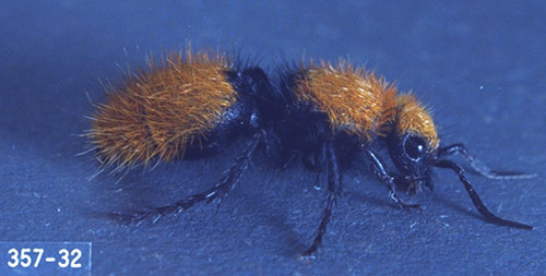
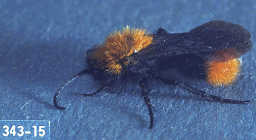
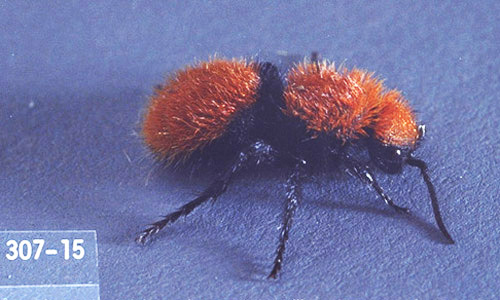
Insects in the family Mutillidae are often referred to as velvet ants because female members of the family lack wings and have coarse setae that cover most of their body, making them resemble hairy ants. To the surprise of most people, mutillids are not ants at all – they are wasps
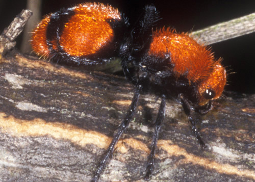
Figure 1. Adult female "cow killer," Dasymutilla occidentalis occidentalis (Linnaeus), a velvet ant. Photograph by Lyle Buss, University of Florida.
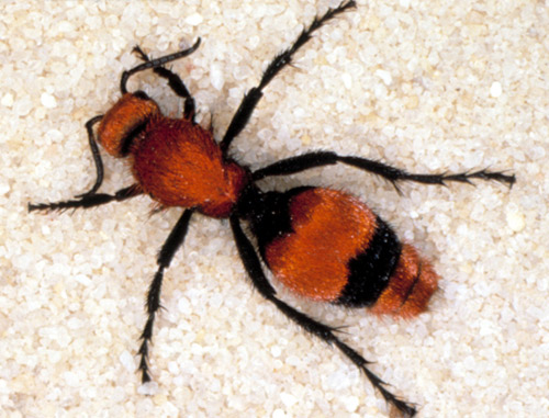
Figure 2. Dorsal view of adult female "cow killer," Dasymutilla occidentalis occidentalis (Linnaeus), a velvet ant. Photograph by James Castner, University of Florida.
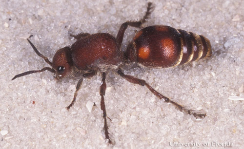
Figure 3. Adult female Dasymutilla sp., a velvet ant. Photograph by Lyle Buss, University of Florida.
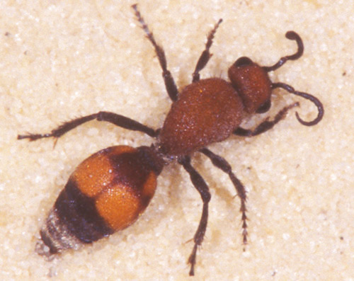
Figure 4. Dorsal view of an adult female Dasymutilla sp., a velvet ant. Photograph by Lyle Buss, University of Florida.
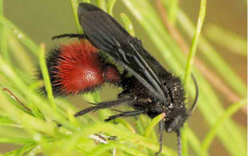
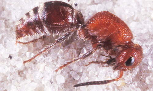
Figure 5. Dorsal view of an adult female Timulla sp., a velvet ant. Photograph by Lyle Buss, University of Florida.
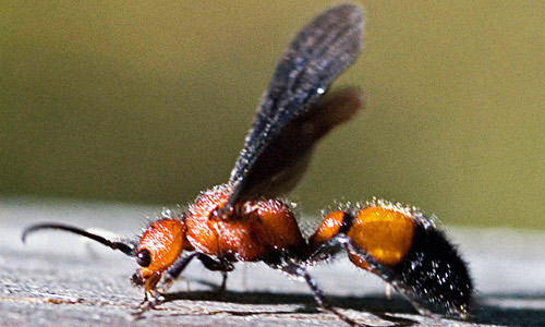
Figure 6. Dorsal view of an adult female velvet ant. Photograph by Clemson University, www.forestryimages.org.
Distribution (Back to Top)
The Mutillidae family contains approximately 230 genera/subgenera and about 8,000 species worldwide (Manley and Pitts 2002). Approximately 435 species occur in mostly arid areas of the southern and western parts of North America (Triplehorn and Johnson 2005). Fifty species in seven genera are found in Florida (Table 1) (Krombein 1979).
Biology (Back to Top)
All Mutillidae are solitary parasitoid wasps that mostly attack mature larvae or pupae of other solitary Hymenoptera. However, velvet ants have been observed targeting non-feeding stages of Diptera, Coleoptera, Lepidoptera, Blattodea, and even some eusocial Hymenoptera (Brothers et al. 2000).
Finding a matching reproductive pair can be difficult, if not impossible for some species, because of the extreme sexual dimorphism displayed within this family. Color patterns and the relative body size of the two sexes of the same species can be very different, making it very difficult to relate one sex with the other (Mickel 1928).
In Florida, male Mutillidae are frequently larger (heavier) than the females. One study in south-central Florida found that in seven out of 13 species, the males were heavier than the females. The remaining six species either showed comparable sizes between the male and female (3) or the female was larger than the male (3) (Deyrup and Manley 1986).
Biological data is very scarce and differences obviously exist between genera and species, especially with regards to their life cycles and development, but the following has been reported for most Mutillidae. Females have the difficult job of locating potential hosts. Suitable hosts may be difficult to find for several reasons including: concealment, low population densities when solitary and lastly, if the hosts are eusocial, they will be heavily defended. Once a host is located, female velvet ants parasitize the "hard" life stages (i.e., hardened pre-pupae, pupae, ootheca, eusocial cells, and cocoons) of the hosts and the emerging velvet ant larvae are essentially ectoparasites of those life stages. (Brothers et al. 2000). Once development is completed, adults leave the nest and seek a mate.
Identification (Back to Top)
All female Mutillidae are wingless and resemble ants. They can easily be differentiated from ants by the lack of petiole nodes, which are present on all ant species. In addition, the mesosomatic (thoracic) segments are completely fused and have at most two segments. The metasoma (abdomen) contains six visible terga (dorsal surface of any body segment) and a "felt line" of dense, closely appressed hairs is located laterally on the second metasomatic tergum (except for the genus Ephuta) (Manley and Pitts 2002, Triplehorn and Johnson 2005).
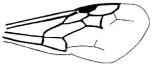
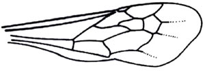
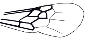
Unlike the female, the male meso- and metathorax are not fused and they have three mesosomatic segments. Their metasomal region contains seven visible terga and a felt line is also on the lateral margin of tergite II. All male Mutillidae found in Florida normally have large wings lacking jugal lobes and forewings with the M and Cu1 veins ending far from the apical margin (Manley and Pitts 2002).
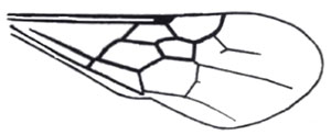
Figure 7. Anterior wing of subfamily Sphaeropthalminae. Note: lacks jugal lobes. (From Manley and Pitts 2002)
Figure 8. Anterior wing, subfamily Mutillinae, with pterostigma membranous or absent. (From Manley and Pitts 2002)
Key to Florida Genera (Back to Top)
The most recently published key to genera and subgenera was issued by Manley and Pitts (Manley and Pitts 2002). Conclusions and pictures contained within this key were derived directly from this publication, but only for genera found in Florida.
Female Specimens (Back to Top)
1. Metasomal segment I completely sessile with second (without petiole) . . . . . 2
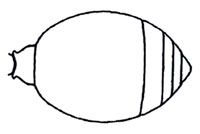
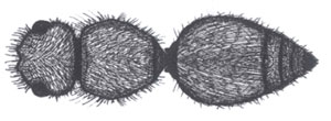
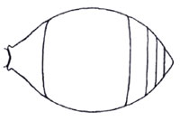
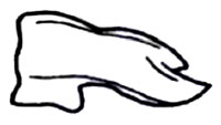
1'. Metasoma petiolate or at most subsessile, with definite constriction between first two segments (Figure 9) . . . . . 5
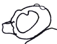
Figure 9. Dorsal view of abdomen with petiolate metasoma. (From Manley and Pitts 2002).
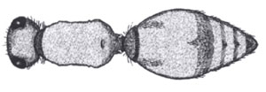
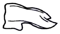
2. Eyes strongly ovate; mesosoma long, rectangular, generally narrowed medially (Figure 10); tergite II generally maculated with two spots or lines of pale setae . . . . . Timulla spp.
2'. Eyes circular to slightly ovate; mesosoma otherwise (short, rectangular or narrowed posteriorly, if narrowed medially, mesosoma pyriform not rectangular); tergite II not maculated with spots of pale setae . . . . . 3
Figure 10. Dorsal view of long, rectangular mesosoma of Timulla spp. (From Manley and Pitts 2002).
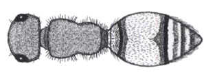
3. Head, in dorsal view, less than twice the width of mesosoma (Figure 11); pygidium usually with lateral carinae, mesosoma narrowed medially; head not dentiform (toothed); if hypostomal teeth present they are not prominent, nor bent apically; posterolateral angles of head, usually not strongly carinate (ridged) . . . . . Pseudomethoca
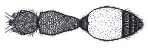
3'. Mesosoma as wide or wider than head in dorsal view, rectangular without fine carina between pronotum and mesonotum (Figure 12); eyes slightly ovate; mandible ventrally toothed or not; sternal felt line absent; plumose setae present or absent; integument visible, usually sparsely covered with appressed and erect simple setae; pygidium variable . . . . . 4
Figure 11. Dorsal view of Pseudometheca simillima (Smith). Note: head less than twice the width of mesosoma. (From Manley and Pitts 2002).
Figure 12. Dorsal view of rectangular mesosoma of Photomorphus spp. Note: mesosoma as wide or wider than head. (From Manley and Pitts 2002).
4. Pygidium dull, shagreened (rough), with parallel carinae only on basal two-thirds or less . . . . . Photomorphus, subgenus Photomorphina
4'. Pygidium smooth and shiny, with complete parallel carinae on disk . . . . . Photomorphus, subgenus Photomorphus
5. Felt lines absent on metasomal tergite II; petiole short, transverse, parallel-sided (Figure 13); eyes distinctly ovate; a band of silvery, dense, pubescence present at apex of petiole and metasomal segment II; small; densely punctuate (subfamily Mutillinae, tribe Ephutini) . . . . . Ephuta
5'. Felt lines present on metasomal tergite II; petiole not transverse or parallel sided; eyes circular to slightly ovate; other characters variable . . . . . 6
Figure 13. Dorsal view of short, parallel-sided petiole of Ephuta spp. (From Manley and Pitts 2002).
6. Plumose setae limited to area of short dense white setae on dorsum of petiole, and apical fringe of tergite II (Figure 14); pygidial area undefined laterally by carinae; genal carina absent; flagellomere II greater than 1.75x length of first; antennal scrobe distinctly carinate dorsally; propodeum elongate . . . . . Sphaeropthalma, subgenus Sphaeropthalma
6'. Plumose setae totally absent . . . . . 7
Figure 14. Dorsal view of apical fringe of tergite II of Sphaeropthalma pensylvanica (Lepeletier). (From Manley and Pitts 2002).
7. Pygidial area well-defined; petiole not diciform (rounded) (Figure 15) . . . . . Dasymutilla
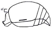
7'. Pygidial area obsolete, not defined laterally; petiole distinctly disciform (Figure 16); Anterior and propodeal spiracles tuberculate (Figure 17) . . . . . Lomachaeta
Figure 15. Dorsal view of non-disciform petiole of Dasymutilla spp. (From Manley and Pitts 2002).
Figure 16. Lateral view of Lomacheata spp. metasoma (af = anterior face). (From Manley and Pitts 2002).
Figure 17. Dorsal view of tuberculate anterior and propodeal spiracles of Lomachaeta variegata. (From Manley and Pitts 2002).
Male Specimens (Back to Top)
1. Compound eye approximately round, not emarginated, smooth and shiny (Figure 18); pterostigma sclerotized . . . . . 2
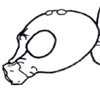
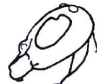
1'. Compound eye with inner margin deeply and sharply emarginated (Figures 19, 20); pterostigma membranous or absent (Figure 21) . . . . . 6
Figure 18. Rounded, not emarginated, compound eye of Photomorphus. (From Manley and Pitts 2002).
Figure 19. Emarginated compound eye of Timulla dubitata (Smith). (From Manley and Pitts 2002).
Figure 20. Emarginated compound eye of Ephuta stenognatha Schuster. (From Manley and Pitts 2002).
Figure 21. Anterior wing with membranous or absent pterostigma. (From Manley and Pitts 2002).
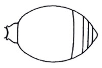
2. Metasomal segment I completely sessile with second (Figure 22); Felt line present on lateral margin of tergite II only; hypostomal tooth not developed; head less than twice width of mesosoma; posterolateral angles of head not strongly carinate or dentiform; wings macropterous . . . . . Pseudomethoca
2'. Metasoma petiolate or at most subsessile, with definite constriction between first two segments (Figure 23) . . . . . 3
Figure 22. Dorsal view of abdomen with non-petiolate metasoma. (From Manley and Pitts 2002).
Figure 23. Dorsal view of abdomen with petiolate metasoma. (From Manley and Pitts 2002).
3. Felt line present on lateral margin of tergite II and sternite II; ventral mandibular tooth present (Figure 24); mesoscutal notauli present (separates mesoscutum into medial and lateral lobes); flagellomere I longer than wide, not like pedicel; hypopygium (last visible abdominal sterum) normal, rounded, lateral margins not carinate or dentiform; mesosternum with pair of longitudinal to transverse dentate ridges anterior to mesocoxae, closer to procoxae; plumose hairs vestigial or absent . . . . . . 6
3'. Felt line present on lateral margin of tergite II only; Mesotibia with two spurs; mesotibia cylindrical, not flattened and arcuate; plumose setae present or absent; mesosternum unmodified . . . . . 4
Figure 24. Mandibular tooth of Photomorphus spp. (From Manley and Pitts 2002).
4. Mandible tridentate apically, broadly emarginated ventrally with small, distinct tooth; eyes and ocelli small, not protuberant, compound eye distinctly separated from mandible, distance from compound eye to posterolateral angle of head distinctly greater than greatest diameter of eye; notauli present, complete; tergites II-V without row of lanceolate (lance-shaped) bristles at distal margin; pterostigma of forewing conspicuous; marginal cell length equal to or slightly longer than stigma; pygidium and hypopygium short, transverse . . . . . Sphaeropthalma, subgenus Sphaeropthalma
4'. Mandible bidentate or tridentate apically, but not emarginate or toothed ventrally . . . . . 5
5. Notauli absent; subplumose setae present; tergites II-V with row of lanceolate bristles at distal margin; pterostigma of forewing vestigial, inconspicuous (Figure 25) . . . . . Lomachaeta
5'. Tergites II-V without row of lanceolate bristles at distal margin; subplumose setae absent; wing venation normal, not greatly reduced; pterostigma of forewing conspicuous (Figure 21) . . . . . Dasymutilla
Figure 25. Inconspicuous forewing pterostigma of the genus Lomachaeta. (From Manley and Pitts 2002).
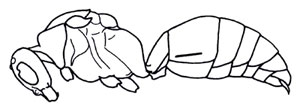
6. Metasomal segment I sessile with second (Figure 26); humeral angles rounded . . . . . Timulla
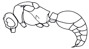
6'. Metasomal segment I slender, short, parallel-sided, not sessile (Figure 27); humeral angles angulate, sharply produced; small; densely punctuate . . . . . Ephuta
Figure 26. Lateral view of habitus with legs removed, Timulla dubitata (Smith). (From Manley and Pitts 2002).
Figure 27. Lateral view of habitus with legs removed, Ephuta stenognatha Schuster. (From Manley and Pitts 2002).
7. Mesocoxae approximate; mandible tridentate apically; mentum never produced into a distinct process . . . . . Photomorphus, subgenus Photomorphina
7'. Mesocoxae separated; mandible bidentate apically (Figure 28); mentum distinctly produced as an anterior tubercle (small nodule) or posterior lingulate (tongue-shaped) process . . . . . Photomorphus, subgenus Photomorphus
Figure 28. Mandibular tooth of Photomorphus spp. (From Manley and Pitts 2002).
Florida Species (Back to Top)
Table 1. List of Mutillidae species reported from Florida
Tribe: Mutillini
Genus: Timulla Ashmead
barbigera barbigera (Bradley)
compressicornis Mickel
dubitata dubitata (Smith)
dubitatiformis Mickel
euphrosyne Mickel
euterpe (Blake)
ferrugata (Fabricius)
floridensis (Blake)
leona (Blake)
ornatipennis (Bradley)
rufosignata (Bradley)
vagans rufinota Mickel
Tribe: Ephutini
Genus: Ephuta Say
battlei battlei Bradley
floridana floridana Schuster
margueritae margueritae Schuster
pauxilla pauxilla Bradley
psephenophila Schuster
puteola (Blake)
sabaliana sabaliana Schuster
slossonae slossonae (Fox)
stenognatha psephenophora Schuster
stenognatha stenognatha Schuster
Subfamily: Sphaeropthalminae
Tribe: Sphaeropthalmini
Genus: Sphaeropthalma
subgenus Sphaeropthalma Blake
pensylvanica floridensis Schuster
pensylvanica pensylvanica (Lepeletier)
Genus: Photomorphus
subgenus Photomorphina Schuster
archboldi (Manley and Deyrup)b
spinci (Bradley)
Genus: Photomorphus
subgenus Photomorphus Viereck
alogus Viereck
banksi (Bradley)
paulus (Bradley)
Tribe: Pseudomethocini
Genus: Pseudomethoca Ashmead
frigida (Smith)
oceola (Blake)
oculata (Banks)
sanbornii aeetis (Fox)
sanbornii sanbornii (Blake)
simillima (Smith)
torrida Krombein
vanduzei Bradley
Tribe: Dasymutillini
Genus: Dasymutilla Ashmead
alesia Banks
asopus bexar (Blake)
asopus cassandra Mickel
castor (Blake)
chattahoochei Bradley
cypris (Blake)
lepeletierii (Fox)
macilenta (Blake)
nigripes (Fabricius)
occidentalis occidentalis (Linnaeus)
pyrrhus (Fox)
rubicunda Bradley
vesta sappho (Fox)
Genus: Lomachaeta Mickel
hicksi Mickel c
aUnless otherwise noted, this list was compiled from the Catalog of Hymenoptera in America north of Mexico (Krombein 1979)
b(Manley and Deyrup 1987a)
c(Pitts and Manley 2004)
Figure 30. Adult female "cow killer," Dasymutilla occidentalis occidentalis (Linnaeus), a velvet ant. Photograph by Lyle Buss, University of Florida.
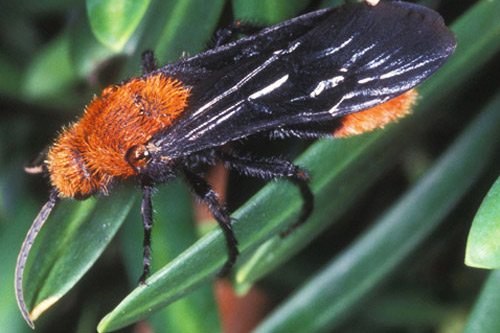
Figure 31. Adult male "cow killer," Dasymutilla occidentalis occidentalis (Linnaeus), a velvet ant. Photograph by Lyle Buss, University of Florida.
Figure 32. Adult male Dasymutilla nigripes (Fabricius), a velvet ant. Photograph by Sean McCann, University of Florida.
Figure 33. Adult male Dasymutilla sp., a velvet ant. Photograph by Lyle Buss, University of Florida.
Figure 34. Adult female Dasymutilla sp, a velvet ant. Photograph by Lyle Buss, University of Florida..
Figure 35. Adult female Dasymutilla sp, a velvet ant. Photograph by Lyle Buss, University of Florida.
Figure 36. Adult female Dasymutilla sp., a velvet ant. Photograph by Ken Gray, Oregon State University.
Figure 37. Adult male Dasymutilla sp., a velvet ant. Photograph by Ken Gray, Oregon State University.
Figure 38. Adult female Dasymutilla sp., a velvet ant. Photograph by Ken Gray, Oregon State University.
Figure 39. Lateral view of an adult female Timulla sp., a velvet ant. Photograph by Lyle Buss, University of Florida.
Figure 40. Adult male Sphaeropthalma pensylvanica (Lepeletier), a velvet ant. Photograph by Charles Schurch Lewallen.
Pest Status and Management (Back to Top)
Mutillidae pose no real threat to humans, livestock, or domesticated animals other than the unfortunate accidental sting associated with unaware victims. Like other stinging insects, only female Mutillidae are capable of stinging. Some species are referred to as "cow killers" or "mule killers," because their sting is said to be painful enough to kill an animal. Despite this assertion, they are not aggressive nor is their venom comparatively toxic. Most female mutillids are so docile, they produce a startle response (an audible stridilation) when provoked to warn potential victims of her sting. The LD50 of their venom when exposed to mice is around 71 mg/kg body weight which is relatively non-toxic compared to the very toxic harvester ant, Pogonomyrmex maricopa, venom (LD50 = 0.12 mg/kg) and the moderately toxic honey bee, Apis mellifera, venom (LD50 = 2.8) (Meyer 1996). No control measures are necessary.
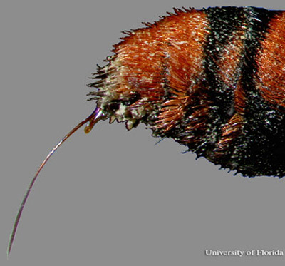
Figure 41. Ovipositor (stinger) of an adult female "cow killer," Dasymutilla occidentalis occidentalis (Linnaeus). Photograph by Jeffrey C. Hertz, University of Florida.
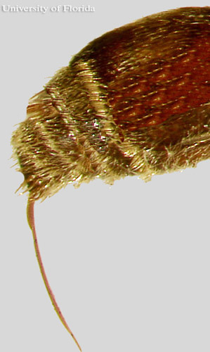
Figure 42. Ovipositor (stinger) of an unidentified, adult female velvet ant, Dasymutilla sp. Photograph by Jeffrey C. Hertz, University of Florida.
Acknowledgments (Back to Top)
I sincerely thank Dr. Don Manley, of Clemson University, for providing me the most current reference literature and taking the time to review this publication. I also thank Dr. Gavin Broad for graciously giving me permission to use numerous images previously published in the Journal of Hymenoptera Research.
Selected References (Back to Top)
- Brothers DJ, Tschuch G, Burger F. 2000. Associations of mutillidae wasps (Hymenoptera, Mutillidae) with eusocial insects. Insectes Sociaux 47: 201-211.
- Deyrup MA, Manley DG. 1986. Sex-biased size variation in velvet ants (Hymenoptera: Mutillidae). Florida Entomologist 69: 327-335.
- Krombein KV, et. al. 1979. Catalog of Hymenoptera in America north of Mexico. Smithsonian Institution Press, Washington.
- Manley DG, Deyrup MA. 1987a. A new species of Dasymutilla (Hymenoptera: Mutillidae) from Florida. Journal of Entomological Science 22: 123-125.
- Manley DG, Deyrup MA. 1987b. A new species of Photomorphus (Hymenoptera: Mutillidae) from Florida. Journal of Entomological Science 22: 57-60.
- Manley DG, Pitts JP. 2002. A key to genera and subgenera of Mutillidae (Hymenoptera) in America north of Mexico with description of a new genus. Journal of Hymenoptera Research 11: 72-100.
- Meyer WL. 1996. Most toxic insect venom. University of Florida Book of Insect Records. (16 December 2013).
- Mickel CE. 1928. Biological and taxonomic investigations on the mutillid wasps. Bulletin of the U.S. National Museum 143: 351.
- Pitts JP, Manley DG. 2004. A revision of Lomachaeta Mickel, with a new species of Smicromutilla Mickel (Hymenoptera: Mutillidae). Zootaxa 474: 1-27.
- Triplehorn CA, Johnson NF. 2005. Family Mutillidae. In Borror and Delong's Introduction to the Study of Insects, 7th ed. Thomson Brooks/Cole, Belmont, CA.
